What is the interpretation of this cardiac telemetry ECG?

Lead I - P-/More R wave
Leads II, III & aVF - P- / like more Q waves
Lead aVR - P+/ More R (instead of more S)
Lead aVL - P+/ R=S (looked ok)
V lead - qr
Interpretation: ectopic atrial rhythm? Dextrocardia? Old inferior MI? Reversed leads?
So, again we checked lead placements and it was correct. After several maneuvers, we finally replaced the whole cable/wire system (not the telebox) and got the correct tracing.

Image 2 - Correct tracing
What happened?
I checked the wires and to my surprise...

Image 3 - Telemetry box connection
Messed-up reversal... I can't imagine that this was possible. Somebody must have cleaned each wires and placed it in this order:
RL (Green) > Chest lead (Brown)
RA (White) > LL (Red)
LA (Black) = ok
LL (Red) > RA (White)
Chest lead (Brown) > RL (Green)
For reference, this is supposed to be the color representation and the normal lead configuration.

Image 4- Normal lead placement/configuration
However, this is what happened here.
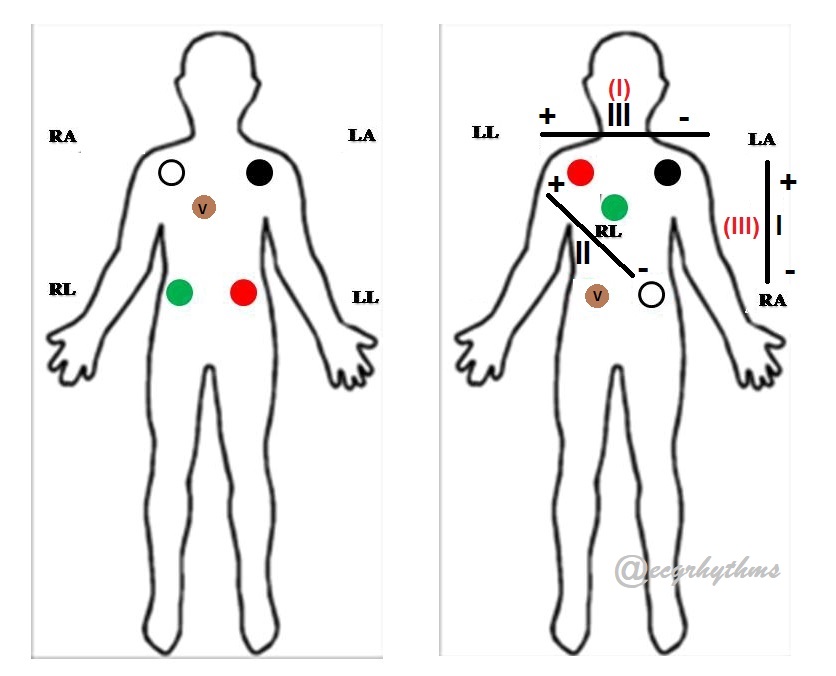
Image 5 - Lead Reversal Diagram
Lead I - LL/LA (instead of RA/LA)
Lead II - became reversed
Lead III - LA/RA (instead of LA/LL)
aVR - LL
aVL - ok
V lead - RL
So, this crazy things are possible... Now we know.
No comments:
Post a Comment
Note: Only a member of this blog may post a comment.