A 70 something patient was admitted due to pneumonia. During routine monitoring this was captured.

Figure 1
The first half of the strip had a ventricular rate of about 94 bpm. After that the RR rate varies at 177 and 188 bpm.
The PP rate also varies. As well as the P wave morphology. The P waves are inverted or negative in II, III and aVF and upright or positive in aVL and aVR. Close inspection of the P wave reveals alternating P wave morphology.
It seemed to have a 2:1 conduction in the first half of the strip and the 1:1 conduction in the latter half. Atrial flutter do not have regular variability as well as atrial tachycardia. So, what is this rhythm?
Few years ago, we have a discussion on this in the EKG Club in FB and here is one of the comments of one of our friends (Dr. Raed).


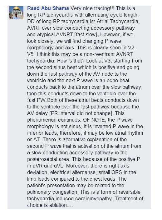

Figure 32 - Proposed ladder diagram
Hopefully this ladder diagram will make this fascinating arrhythmia easier to understand. So, our thought was that this could be a low atrial rhythm that reentered and created an atrial echo. So, this can explain the 2 different P wave morphology and rate. On the latter part of strip, after the atrial echo, the impulse was conducted to the ventricle. This created the regular variable RR interval.
So, what happened to the patient? This event happened twice (about 15 and 20 minutes). The patient was asymptomatic and the tachyarrhythmia was self-terminating. The patient improved with respiratory issue and was discharged after a few days. The tachyarrhythmia did not recur during this admission.
#77
No comments:
Post a Comment
Note: Only a member of this blog may post a comment.