1/28/2016

De Winter's ST/T ECG changes in huge anterior wall MI

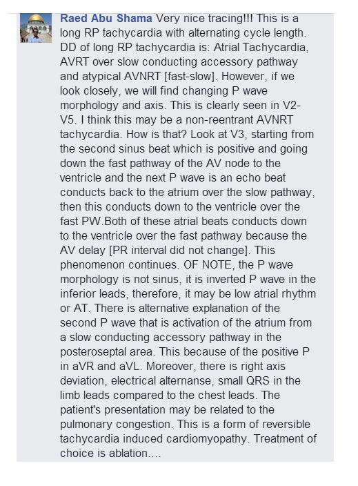
(Case post by Dr. Bojana Uzelac)
This is ECG from 70yo man complaining of strong chest pain, started 20 minutes ago. He is pale, sweating and looking weak. His vitals are: HR: 60/min, RR: 18/min, TA: 110/70mmHg, Sat O2: 96% on the room air.
What is your ECG interpretation?
How would you manage this patient?
(Courtesy of Dr. Ivana Kundačina)
Originally posted in:
Conclusion:
Thank you all for your answers. I’m so glad that almost all readers were able to recognize this ECG pattern.
This was a case of de Winter T/ST ECG changes representing a huge anterior MI.
De Winter T/ST pattern is relatively new ECG finding associated with ACS and LAD occlusion. Instead of STEs in precordial leads, some patients with AWMI may show 1-3mm up-sloping ST-segment depression at the J point in leads V1-V6 that continued into tall, positive symmetrical T waves. This ECG pattern was first reported by de Winter et al.(N Engl J Med 2008; 359:2071-2073). According to them, this finding was observed in about 2% of AWMI.
Image 1. Features from de Winter article
It was notice, that despite early and successfully LAD opening, the loss of myocardium was significant.
Some authors have suggested that the de Winter pattern should be considered a “STEMI equivalent”. (Rokos I, et al. Appropriate Cardiac Cath Lab activation: Optimizing electrocardiogram interpretation and clinical decision-making for acute ST-elevation myocardial infarction 2010; 160(6):995–1003). They’ve proposed that patients with chest pain and this ECG feature should receive prompt reperfusion therapy.
There is a difference between early, hyperacute T waves and de Winter T waves. Hyperacute T waves are transient feature, seen in the early stage of AMI and evolve into STEs. De Winters T/ST pattern, however, is static and persistent form. It was on average recorded 1,5 hours after chest pain onset. The electrophysiological explanation of this pattern remains elusive, with several theoretically possible reasons.


Image 2. ST segment associated with hyperacute T waves may be elevated or isoelectric; ST segment associated with de Winter T waves shows up-sloping STD at the J point.
Some people may mistake another two ECG features: de Winter and hyperkalemia T waves. But, there are differences in both clinical presentation and ECG findings. In hyperkalemia, T waves are much more narrow and tented; usually without preceding up-sloping ST segment depression. T waves in de Winter pattern are on broad basis, symmetrical and associated with upsloping STDs.
Image 3. Difference between de Winter T/ST changes and hyperkalemia
So, what happened with our patient? I was consulted about this ECG, and we decided to activate the cath lab. During transportation this patient looked even worse. Another ECG was recorded 20 minutes after first one (I have only precordial leads available). It shows further slowing of heart rate with ventricular bigeminy.
Image 4. Precordial leads from second pre-hospital ECG.
This is the hospital ECG, recorded about 30 minutes after the first one.
Image 5. Hospital ECG: de Winter T/ST evolved into anterior STEMI
Now there is huge antero-lateral STEMI, with R wave lost in precordial leads and small QRS amplitudes.
Angiography found 99% occluded proximal LAD; successfully opened and stented. But, despite that, he deteriorated rapidly. His systolic BP dropped to 60 and he became altered mental status. He was intubated and put on vasopressor support. He required intra-aortic balloon pump, as well, due to severe cardiogenic shock.
After interventions patient was hospitalized in ICC unit and another ECG was done.


Image 6. ECG after LAD opening
There are significant Q waves in all precordial leads, with a less amount of STEs and terminal inversion of T waves.
Image 7. Comparison between pre- and after procedural ECGs
Despite reperfusion, his prognosis was challenging. The troponin I came elevated, 0,276. Others lab analysis were unremarkable.
Luckily, this patient reacted well on therapy and improved. He was extubated next morning and remained on intra-aortic balloon pump for next 24h. On inotropic support his BP was 110/80 and he got good diuresis. He survived this huge STEMI.
De Winter ECG findings may be subtle and difficult to notice; therefore it is very important that all emergency providers be aware of this ECG pattern.

1/24/2016

Sudden PR interval prolongation due to concealed conduction

 
Figure 1 - ECG case
 
An adult patient admitted for altered mental state.
What is the probable reason of the prolongation of the PRI (red
arrow)?
 
Answer: Concealed retrograde conduction of a premature junctional beat delaying AV conduction (prolonging a PR interval)
 
 
Figure 2 - ladder diagram

#321

1/23/2016

Beware of VTactitis


Beware of artifacts simulating VT/VF. Machine even read the rate at 266 bpm. However, organized QRS can be seen in III. You might end up hangning your amiodarone or shocking a patient. :) Try evaluating telemetry tracings in full disclosure.

#629

Sinus rhythm, 2:1 AV block due to Mobitz II


This is a 70 yo pt who was admitted due to weakness. What is your interpretation?


Figure 1 - ECG case

Sinus rhythm, 2:1 AV block due to Mobitz II

Figure 2 - case marked

The sinus (atrial) rate is about 88 bpm and the ventricular rate is about  42 bpm. The QRS has right bundle branch block (RBBB) configuration. There are 2 P's for every 1 QRS. This is termed 2:1 AV block. A 2:1 AV block is a special form of second degree AV block. It can either be second degee type I (Mobitz I or Wenckebach) or second degree type II (Mobitz II). A longer strip is needed to elucidate the mechanism. In this case, this 10 second strip captured 3:2 AV conduction showing a dropped beat with no PR interval prolongation prior to the dropped beat. So, this is second degree AV block type II (Mobitz II) manifesting as 2:1 (predominantly) and 3:2 AV conduction.

Sinus rhythm, advanced heart block

Figure 3 - Advanced Heart Block

This 10 sec strips is minutres after the previous strip. QRS #2 is a conducted beat and after that there is AV dissociation due to accelerated idioventricular rhythm (AIVR). Obvious P waves are marked in read and hidden P waves are marked in green. When the AV conduction ratio s 3:1 or higher, the rhythm is called advanced AV block. This block occurs because of AV node or His-purkijne disease and not retrograde concealment in the AV node or His-Purkije system caused by junctional or ventricular escape complexes. A few minutes later, 2:1 AV blocked recurred (not shown). A permanent pacemaker was eventually placed.

Reference:

Surawicz B and Knilans TK. 2008. Chou’s Electrocardiography in Clinical Practice. 6th ed. PA. Saunders-Elseiver

Issa Z, Miller J and Zipes D. 2012. Clinical Arrhythmology and Electrophysiology A Companion to Braunwald’s Heart Disease 2nd Ed. PA Saunders

Das and Zipes. 2012. Electrocardiography of arrhythmias : a comprehensive review. Elsevier PA

#628

1/19/2016

A long RP tachycardia with alternating cycle length


A 70 something patient was admitted due to pneumonia. During routine monitoring this was captured.


Figure 1

The first half of the strip had a ventricular rate of about 94 bpm. After that the RR rate varies at 177 and 188 bpm. 

The PP rate also varies. As well as the P wave morphology. The P waves are inverted or negative in II, III and aVF and upright or positive in aVL and aVR. Close inspection of the P wave reveals alternating P wave morphology. 

It seemed to have a 2:1 conduction in the first half of the strip and the 1:1 conduction in the latter half. Atrial flutter do not have regular variability as well as atrial tachycardia. So, what is this rhythm?

Few years ago, we have a discussion on this in the EKG Club in FB and here is one of the comments of one of our friends (Dr. Raed).


Figure 32 - Proposed ladder diagram

Hopefully this ladder diagram will make this fascinating arrhythmia easier to understand. So, our thought was that this could be a low atrial rhythm that reentered and created an atrial echo. So, this can explain the 2 different P wave morphology and rate. On the latter part of strip, after the atrial echo, the impulse was conducted to the ventricle. This created the regular variable RR interval. 

So, what happened to the patient? This event happened twice (about 15 and 20 minutes). The patient was asymptomatic and the tachyarrhythmia was self-terminating. The patient improved with respiratory issue and was discharged after a few days. The tachyarrhythmia did not recur during this admission.

#77

1/18/2016

Atrial fibrillation with entrance block, nonparoxysmal junctional tachycardia with type I (Wennckebach) exit block


Figure 1 - Lead II - rhythm strip 

This is atrial fibrillation with entrance block, nonparoxysmal junctional tachycardia with type I (Wennckebach) exit block


Figure 2 - Ladder diagram

There are no discernible P waves. You might be misled that there are distortions in the terminal portions of the T waves but they are not consistent and you cannot march them out. So, there is atrial fibrillation (AF).

AF, however, cannot generate regularly occurring patterns. Thus, there is a AF with entrance block. Entrance block denotes failure of an impulse to reach, enter, suppress, reset, or discharge a dominant pacemaker.The next dominant pacemaker that can generate a regular pattern is the AV junction. However some of the impulse from the AV junction is blocked (exit) and this creates a pattern/group-beating. Group-beating is marker of a Wenckebach periodiocity.

To give us an idea of the AV junction rate, we will use the 3:2 pattern or the 2 QRS and 3 junctional beats. The RR interval of R2 to R4 is about 1320 ms. So, we divide 1320/3 and we get 440 ms as the interectopic interval or a rate of about 136 bpm ( small squared method = 1500 / (440/40) ) for the AV junction or there is junctional tachycardia. Because this pattern is persistent then this is non-paroxysmal junctional tachycardia.

In the ladder diagram, the AF is blocked and the approximate origin of the junctional beats is set at an interectopic interval of about 440 ms (~11 small squares).There is a 3:2 and 2:1 pattern as shown in the ladder diagram.


#627 

Pseudo-first degree AV block


Figure 1 - 30 sec continuous strip

This looked like sinus bradycardia (~44 bpm) with a very long PR interval (800 ms or 0.80 sec) or first degree AV block.


Figure 2 - 30 sec continuous strip few minutes prior to the strip in Figure 1

In #1, a distinct P wave can be seen in R2 but you will note that it merged with the QRS. Because of the merging, the shape of the QRS is distorted. In #2 and #3, the P wvave can be seen after the QRS. On the last QRS complex, the P wave is seen farther awway from the QRS. The QRS are junctional complexes. So, there is AV dissociation. The rate of the sinus beat is almost the same with the junctional rate. This is called isorrhythmic AV dissociation.

The ECG strip in Figure 1 is not a true first degree AV block but a pseudo-first degree AV block. The strips are sinus bradycardia with junctional beats creating AV dissociation (isorhythmic AV dissociation)

After a few hours (of stopping a beta-blocker), the sinus rate increased to about 60 and captured the ventricles. 

#626

1/17/2016

Hyperkalemia


An 80yo with history of HTN, dyslipidemia, CAD, s/p CABG on lisinopril, furosemide, potassium and spironolactone was admitted due to syncope. BP  systolic 70's PR 80 RR 18 and O2 sat 95 at 5L O2. Patient is lethargic with dry oral mucosa, clear breath sounds, regular cardiac rhythm and no murmur, flat, soft abdomen, no pedal edema.This is the ECG.


Figure 1 - 12L ECG

The 12L ECG revealed a regular wide QRS rhythm, tall T waves and the P waves are difficult to discern. The QRS and T wave segment seemed to merge or having a "sine" wave pattern. 

Laboratory work-up revealed creatinine of 3, BUN 62, bicarb 12 and K of 8. Patient was hydrated and NaHCO3, D50 and insulin, kayexalate were given. So, this patient has hyperkalemia from acute renal failure probable from diuretics (and patient was on ACEI and K supplement)

Progressive hyperkalemia produce distinctive sequence of events affecting the QRS (depolarization) and ST=T segments (repolarization). The normal serum potassium is between 3.5 and 5 mEq/L. The changes would be narrowing and peakng of the T waves ("tented" and "pinched" shape). Further potassium elevation will make P waves small and may disappear entirely. Continued elevation will produce intraventricular conduction delay (widening of the QRS), sine-wave pattern and asystole.

Reference:

Goldberger A. 2013. Goldberger’s Clinical Electrocardiography : A Simplified Approach 8Ed. Ph Elsevier

#307

1/13/2016

Retrograde Wenckebach


Sinus rhythm with concealed intranodal reentry and an episode of accelerated junctional rhythm with retrograde Wenckebach

Edit: (092823)

I would like to thank Dr. @NaokiThukishima who was able to point-out that after R4 is a PAC and not a retrograde P wave. Thus this arrhythmia was initiated by PAC leading to AVNRT. Dr. @AThomazAndrade also suggested that this could be AVNRT with a Wenckebach. Thus, the proposed ladder diagram is shown below (top diagram).


References:

Fisch C and Knoebel SB. 2000. Electrocardiography of Clinical Arrhythmia. New York. Futura Publishing Co.

Wang K.2013. Altas of Electrocardiography. India Jaypee Bros

#625

1/11/2016

Concealed retrograde conduction


Sinus bradycardia with blocked/non-conducted sinus beats due to concealed retrograde conduction of PVC's (multifocal)

#624

1/10/2016

QRST alternans


A 60 yo with a h/o of DM and chronic kidney disease is admitted due to respiratory failure and shock. This ECG was noted while being monitored?


Figure 1

This was refered to cardiology service because of QRS alternans. Cardiac tamponade as a cause of the QRS alternans was ruled-out by echocardiography.

On the 12 L, there is sinus rhtyhm. On the QRS with normal duration, there is diffuse flattening of the ST segment and the extra distortion could be U waves. Seeing that, the QT is prolonged. On the alternating wide QRS complexes, the PR interval are short and NOT CONSTANT. The T waves are pronounced and inverted. Thus this is thought to be fused PVC's in bigeminal pattern creating pseudo-alternans. 

This patient was noted to have hypokalemia. It was replaced and the bigeminal pattern was resolved.


#299 

Bradycardia due to a neuro device


A young patient came in due to seizure. What is happening here? Is there a device malfunction?


Figure 1

No wires are going to any chamber. Instead, the device was a vagus nerve stimulator (VNS).  This is the tracing (Figure 1) when the device was being adjusted for the optimal output until no bradyarrhythmic events were noted.


Figure 2

Patient had this device (VNS) for a number of years. During this admission, bradyarrhythmic events  (Figure 2) were noted. Device behavior was observed. No bradycardic events were noted when device was off but had several bradycardic events when it was turned on (~ 3 seconds - not shown here) So, neurology service had to adjust the output.

#124

1/09/2016

STEMI : Anterior wall

An adult pt with a history of HTN came in due to chest pain with radiation to the left arm associated with dyspnea. No nausea or vomiting. VS 140/80 afebrile HR 80’s RR 20 and sat 98% at room air. No JVD, CBS.


Figure 1

ECG showed SR in the 90’s with ST elevations (STE) in V1-V5 (max STE in V2), hyperacute T waves prominent in V2/V3 and ST depressions (STD) in III, aVF and II. QTc is 498 ms.

Intervention revealed occlusion in the left anterior descending (LAD) artery. Stent was then placed.
Maximum troponin was 1.5 and echo showed and EF in the 60’s. Patient was then discharged after a few days.

#280


Isorhythmic AV dissociation


Figure 1 - Long lead II and V1

What is the rhythm?

This is regular narrow QRS complex rhythm (~88  bpm). Most would call this accelerated junctional rhythm.


Figure 2 - Full disclosure with P waves marked

P waves are marked in red arrows. They are upright in II and negative in aVR. This means that there still sinus rhythm. However, the ventricles is controlled by the junction. There is AV dissociation. When the atria and ventricles are dissociated and the rates are the same, this is called isorhythmic AV dissociation. 


Figure 3 - Follow-up strip 

Follow-up strip shows the separation and merging of the P and the QRS.

These are strips (static mode)  are difficult to interpret or often misled an interpreter. Longer strips like reviewing them on telemetry will make the interpretation easier. 


#278

Right Ventricular Outflow Tract Ventricular Tachycardia


Figure 1 - Remote Monitoring


Figure 2 - 12 L lead ECG

Vignette: A 45 yo pt c/o of palpitations with normal EF

The strip shows regular wide QRS tachycardia (~214 bpm). in the 12 lead,  it shows left bundle branch (LBBB) morphology, inferior axis, late precordial R wave transition, positive QRS complexes in the inferior leads and wide notched R waves in I and aVL. Electrophysiological study showed right ventricular outflow tract VT and ablation was done.

Ventricular  tachycardia in patients with normal hearts is called idiopathic VT. There are 2 types - adenosine sensitive idiopathic VT and verapamil sensitive fascicular VT.

The most common form of idiopathic VT originates from the VT outflow tract. Seventy-five to eighty percent originate from the right ventricular outflow tract (RVOT) and the rest can originate anywhere in the ventricles.

RVOT VT have LBBB morphology with precordial R wave transition in lead V3 or V4 and positive QRS complexes in the inferior leads. RVOT can be divided into septal anterior, posterior and lateral walls (Figure 3). 


Figure 3 - Localization of Vt origin by QRS morphology

*Precordial transition - QRS transition in precordial leads with change where R wave becomes greater than S wave. Transition at or beyond lead V4 means late transition.

Reference:


#623

Use all available leads in rhythm interpretation


Figure 1

A adult patient is admitted due to GI bleed.

What is the interpretation?


Figure 2

The rhythm is SR with long PR interval (first degree AV block), right bundle branch block (RBBB) and showing Wenckebach periodicity (aka Mobitz I) with 4:3 AV conduction.  If you rely only lead II, which is usually the monitoring lead. You will probably scratch your head. You have to utilize all available leads. Distinct P waves are marked with red arrow. The non-conducted beats are marked with black arrows.

The patient was asymptomatic and none was done for regarding the rhythm.

#274

1/08/2016

Hypokalemia


A 40 yo F patient with h/o of alcoholism is admitted due to abdominal pain and vomiting.


Figure 1

There is a baseline artifacts (part of real-life ecg's). This is a regular narrow complex tachycarduia with prolonged QT/QTc. A QT interval that crosses the middle of an R to R is highly suspicious for for prolonged QTc. The QT interval is about 400 ms and at a rate of about 100-107 bpm, the QTc is  516-534 ms which is prolonged. There is also diffuse ST sagging. These ECG findings can be seen in hypokalemia. In this case, the K was 2.5. The tachycardia was due to alcohol withdrawal.

#273 


1/03/2016

Wide QRS Tachycardia


An adult patient came in due to palpitations. What is the rhythm?


Figure 1

What is a Wide Complex Tachycardia?

A wide complex tachycardia (WCT) is defined as a cardiac rhythm with  a rate ≥ 100 bpm and QRS width/duration 120 ms or 0.12 sec. Other terms used aside from WCT is wide QRS tachycardia or WQRST. 

A WCT is also defined by the V1 morphology. So, a WCT can have a right bundle branch block (RBBB) configuration or left bundle branch block configuration. A RBBB is recognized by a QRS duration  120 ms with a predominantly positive portion in V1. LBBB has a QRS duration of 120 ms with a predominantly negative  terminal portion in V1.


WCT can be:

Ventricular Tachycardia (VT)

Supraventricular tachycardia (SVT):
A) with aberrancy in the His-Purkinje system
B) with anterograde accessory pathway conduction
C) with bizarre baseline QRS
D) in presence of drug effect or electrolyte imbalance

Ventricular pacing

Electrocardiogram artefact

VT requires the participation of structures below the bundle of His while SVT requires the participation of structures above the bundle of His.

Why take the time to differentiate WCT?

The purpose of arriving at the correct diagnosis is to avoid harm to the patient. If SVT is treated as VT and given amiodarone or electrical cardioversion may not be harmful but not the optimal therapy. If it was atrial flutter, cardioversion will entail a risk of stroke.  If VT is treated as SVT (using diltiazem/verapamil), hemodynamic deterioration may occur. If SVT are managed as VT, they might be placed on long-term amiodarone which carries a number or long-term problems. We should also not fall into the trap that stable patients (minimal symptoms) with WCT have SVT or unstable patients with WCT are VT. Also, we should not fall into the belief that a WCT terminated by adenosine or verapamil is SVT because some VT are sensitive to these drugs.

So, we go back to the patient and the long time dictum to treat the patient and not the monitor. If the patient is unstable then immediate cardioversion and then if the patient is stable then the various algorithms are used.


Table 1 Predictive Values and Accuracies of the Most Common Ventricular Tachycardia Criteria

Others would just say treat a WCT as VT because by prevalence alone we will be correct 4 out 5 times because pre-test probability that a WCT being VT is in excess of 80%. However, it will defeat the reason of having warm thinking body and avoiding harm to our patients.

As we can observe, most algorithms differentiating VT from SVT with aberrancy focus on characteristics unique to VT. If those characteristics are not present, then it is presumed SVT until proven. Also, we should recognize that algorithms find it hard to distinguish VT from pre-excited SVT.

VT description

In SVT, the relationship to the R the P is used to classify into 2 big groups (short RP or long RP SVT). In VT, the QRS morphology is used. It could either be RBBB WCT or LBBB WCT. This is done by observing the terminal deflection in V1. If the terminal deflection is negative then it is LBBB and if the terminal deflection is positive then it is RBBB WCT.

Here is a very educational graphic from Drs. Garner J and Miller J. 


Figure 2 - Morphological Criteria Discriminating VT from SVT from Garner J and Miller J.

Using the different criteria for the case

  • Brugada algorithm - RS interval > 100 ms (120 ms in V5 and V6) = VT


Figure 3 - RS interval > 100 ms

  • Morphologycal criteria for VT - 


V1-  BIphasic QRS - QR
V6 - R:S ratio < 1


Figure 4 - V1 and V6

  • aVR algorithm (aka Vereckei) - Vi/Vt in V6 0.1/0.4 < 1 = VT


  • Bayesian approach - right superior axis, qR in V1 = VT


  • RWPT criteria - V1 50 ms = VT


Back to the case

This is a regular wide QRS monomorphic tachycardia with right bundle branch (RBBB) morphology and right superior axis at cycle length 400 ms (~ 150 bmp).

This is sustained monomorphic ventricular tachycardia, emanating from the left ventricular focus. Amiodarone was given terminating the tachycardia. It was also found out the EF was low and AICD was offered.

References:

Brugada et al. 1991. A New Approach to the Differential Dx of a Regular Tachycardia with a wide QRS Complex.Circ 83:1649-1659 - http://circ.ahajournals.org/content/83/5/1649.full.pdf+html

Bonnow et al. 2014. Braunwald's Heart Disease: A Textbook of Cardiovascular Medicine. 10th Edition. PA.Saunders 

Drew B and Scheinman M. 1995. ECG Criteria to Distinguish Between Aberrantly Conducted Supraventricular Tachycardia and Ventricular Tachycardia: Practical Aspects for the Immediate Care Setting. PACE 18:2194-2208


Garner J and Miller J. 2013. Wide Complex Tachycardia – Ventricular Tachycardia or Not Ventricular Tachycardia, That Remains the Question. Arrhythmia & Electrophysiology Review 2(1):23–29


Miller et al. 2006. The Value of 12-Lead ECG in Wide QRS Tachycardia Cardiology Clinics 24:439-451


Sandle and Marriot.1965.The Differential Morphology of AnomalousVentricular Complexes of RBBB-Type in Lead V1 Ventricular Ectopy versus Aberration. Circulation 31: 551-556

Stahmer SA and Cowan R. 2006. Tachydysrhythmias. Emergency Medicine Clinics of North America 24:11-40


#268